Everything you need to know about getting a 20 week ultrasound. Plus, what exactly are sonographers looking for during the scan? Find out here!
The 20-week milestone is a big one! Leaving those early pregnancy signs in the dust, you’re at the halfway point of your pregnancy. ? This is also the time when many women get a 20 week ultrasound.
In this article, we’ll explain everything you need to know about the 20 week ultrasound, including:
- What healthcare providers look at during the 20 week ultrasound
- What to expect at your 20 week ultrasound
- The pros and cons of having a 20 week ultrasound
- Plus, how to minimize the effects of an ultrasound
What is the 20 Week Ultrasound?
The 20 week ultrasound, also known as the anatomy scan, is when a sonographer uses an ultrasound machine to:
- Check for physical abnormalities in baby
- Check mama’s uterus, fluid levels, and placenta
- Determine baby’s sex
Though it’s referred to as the 20 week ultrasound, most women have the exam some time between 18 and 21 weeks.
20 Week Ultrasound: How to Prepare
The preparation is minimal for the scan, but here are a few do’s and don’t’s:
DO
- Wear lose, comfortable clothing. Elastic pants or a flowing dress are great options since you can easily access belly for the scan.
- Get childcare for older siblings. Many radiology centers do not allow children to accompany mamas to the appointment.
DON’T
- Empty your bladder. To ensure the inside of the abdominal area is clearly seen on the ultrasound images, you should have ample fluid in your bladder. Many practitioners will ask you to drink at least 16 ounces, one hour before test.
Pregnant? Get my FREE week-by-week updates! – Week by Week Promo [In-article]
Track your baby’s growth, find safe and natural remedies, and have fun along the way!
Get Pregnancy Updates!
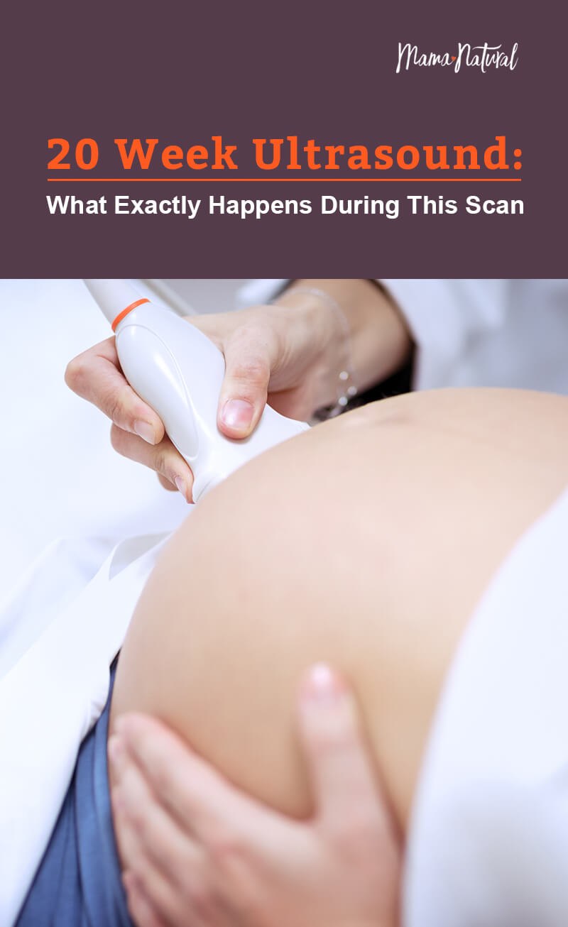
20 Week Ultrasound: What Happens
Here’s what you can expect at your 20 week ultrasound:
- You will be asked to remove clothing so the sonographer can easily access your bare belly.
- You will then lay semi-reclined on an exam table with belly exposed from the lower ribs to the top of the hips.
- The sonographer will spread a clear gel on your belly.
- Next, the sonographer will move a probe called a transducer all over your abdomen. This transmits sound waves that go through the abdominal wall. These waves bounce off of the organs and bones of your developing baby to produce images on the monitor.
- During this process, the sonographer looks at a screen that shows the image of the baby, records measurements, and takes pictures. Oftentimes, this screen is also visible to you and others who are present at the ultrasound.
- There is a lot of information to gather during the 20 week ultrasound. Expect to be there anywhere from 30 minutes to an hour, depending on how wiggly your baby is and how fast your sonographer is.
- If the sonographer is tight-lipped throughout the process, don’t fret. Sonographers need to focus on getting the information they need. They’re also not legally supposed to share information with you. That being said, most sonographers will point out some obvious things he/she is seeing, like the beating heart and spine, and baby’s sex (if you are interested to find out if you’re having a boy or girl)!
- Once the sonographer has finished the ultrasound, they will most likely snap some pictures of your baby for your records—and for you to take home, if you wish.
- Once the ultrasound is complete, a radiologist will review your results, then forward them to your healthcare provider, who will share the results with you.
(Note: It’s always a good idea to ask your healthcare provider if there are any special procedures that you should follow.)
20 Week Ultrasound: What are Health Providers Looking For?
So now you know what to expect, but what exactly are they looking for? A 20-week ultrasound provides a fairly comprehensive look at what’s going on in your uterus!
Here’s the full scope:
-
- Heartbeat: The sonographer will look for a heartbeat that has a nice variability, taking note of its rhythm. According to studies, fluctuations of beat-to-beat intervals is considered the most important fetal heart rate indicator of baby’s development. In a relaxed state, the variation between heart beats is high. In a stressed state, the variation is low. A high heartbeat variation shows resilience and correlates with overall health. (source)
- Amniotic fluid: The sonographer will measure the pockets of fluid that surrounds the baby to make sure there is an average amount of fluid– not too little, not too much, an indirect sign of fetal health.
- Baby’s position: Next, they take a look at how the baby is positioned in the uterus—head up or head down! (Note: At this point in the pregnancy, a baby has tons of room—so don’t fret if baby is breech, there is plenty of time for baby to get into an optimal position.)
- Anatomy measurements: The sonographer takes different measurements, like head circumference, femur length, and the abdominal circumference. These measurements help the sonographer:
- Give your baby a percentile for size.
- See if baby’s size correlates with the due date. Occasionally, if the baby is larger or smaller than expected, your due date may be changed.
- Anatomy development: After the measurements, the sonographer conduct’s a head-to-toe exam on baby, looking at the spine, brain, heart, lungs, diaphragm, stomach, intestines, kidneys, and bladder for any abnormalities. They even count the fingers and toes!
- Baby’s sex: If you so choose, the sonographer can reveal baby’s sex!
- Placenta: The next part of the scan involves locating the placenta to determine it’s position and check for a condition called placenta previa. He/she will also examine overall placenta health and vascularity.
- Umbilical cord: Then, the sonographer looks at the umbilical cord to see where it attaches to the placenta (and the baby!). He/she also notes blood flow in the placenta and the cord.
- Maternal structures: Lastly, the sonographer will take a peek at some of the other maternal structures like:
- The uterus to check for fibroids.
- The ovaries to check for cysts or masses.
- And the cervix to measure length and position.
What Do They Screen For During the 20 Week Ultrasound?
Through the above process, this detailed scan allows the sonographer to look specifically for indications of 11 rare, but serious conditions, including:
While this scan can give you quite a bit of information about your baby’s well-being, it is important to remember that baby is still developing. Please know that a positive 20 week ultrasound is a great milestone—and one that should be celebrated—but it can’t pick up everything.
Is a 20 Week Ultrasound Necessary?
One might get ‘mandatory’ and ‘routine’ confused when it comes to prenatal testing. Though the 20 week ultrasound is routine, it is not necessarily mandatory.
Not every woman wants or even needs a 20 week ultrasound.
In fact, studies do not show improved baby or maternal outcome in those who opt to have a mid-pregnancy ultrasound.
That said, many mamas and papas prefer to have a 20 week ultrasound to bond with the baby and get peace of mind.
What if the 20 Week Ultrasound Indicates a Problem?
Identifying a problem during the 20 week ultrasound may or may not be a good argument for having the scan to begin with. You’ll have plenty of time to understand the condition and set up the appropriate medical team for after-birth care.
Keep in mind, though, some conditions uncovered during a 20 week ultrasound resolve themselves with time, such as:
- Water on the kidneys. Sometimes a scan will show water on the kidneys, which most often clears up on its own as baby continues to grow and develop.
- Placenta previa. Another common scenario is when a scan identifies placenta previa. According to studies, 98 percent of marginal placenta previas are resolved at a mean gestational age of 28.6 weeks.
Once an ultrasound indicates a problem, your healthcare provider may suggest further scans and follow-up to tests to track progress and gather more information.
Is the 20 Week Ultrasound Dangerous?
Though ultrasounds are regarded as safe to mama and her baby, experts say, like any medical procedure, it does carry some inherent risk—namely the potential for misdiagnosis and the fact that ultrasound does impact the body. Current research is not strong enough to prove ultrasound harms mama or her baby, but because it’s a form of energy that heats tissue, it may have an effect on the biological tissues it traverses. (source)
Bottom line: Many women have ultrasounds every day, with no known effects. Still, more research is needed.
Just remember: It’s recommended that ultrasound exams be performed only for medical reasons by qualified health care professionals.
When Are Ultrasounds Necessary?
As natural mamas, we try to avoid as many interventions as possible. But sometimes it’s in your best interest to have an ultrasound. Here are few of those scenarios:
- Monitoring twins or higher order multiples
- Understanding why you’re measuring large/small
- Identifying the reason for vaginal bleeding in pregnancy
- Confirming position of placenta if placenta previa was an issue
How to Minimize the Effects of Ultrasounds
Because there is still much to learn about the safety of ultrasounds, consider these strategies to reduce the effects of ultrasound:
- Stick with a more traditional 2D ultrasound. Skip the 3D or 4D. This fad of having a more lifelike picture of your baby in the womb is on the rise, despite warnings from the Food and Drug Administration (FDA) and the American Congress of Obstetricians and Gynecologist (ACOG).
- See if you can wait until 22 or 23 weeks. This gives your baby a little more time to develop and the placenta a chance to move. Sometimes, when you opt for having the scan on the earlier side, doctors or midwives may need to do a repeat scan in just a few weeks to better assess the baby’s development.
- Ask to forgo the doppler and use a fetoscope instead. At around 20 weeks, you can listen to baby’s heartbeat via a fetoscope instead of a hand-held doppler. Studies show that a handheld doppler emits waves at a higher temperature. Though there is no conclusive evidence, as stated above, this could potentially affect the tissue it transverses.
- Forgo pictures. A sonographer may spend extra time getting a perfect profile pic, and extra time means the potential for extra exposure.
- Be upfront with the sonographer. It’s ok to let the sonographer know that you are concerned about the effects of ultrasound. Ask for the scan to be complete, but efficient.
How About You?
Did you have a 20 week ultrasound? Is there anything you wish you knew beforehand?